Popular culture frequently conflates celiac disease with lifestyle dietary choices, mild food allergies, or general digestive sensitivities. In clinical reality, celiac disease is a severe, systemic autoimmune condition. For individuals with the genetic predisposition, consuming even microscopic amounts of gluten—a protein composite found in wheat, barley, rye, and their derivatives—sets off an aggressive internal response. Rather than a simple allergic reaction that manifests as hives or immediate gastrointestinal distress, celiac disease launches a targeted immunological assault against the individual’s own tissues, specifically the mucosal lining of the small intestine.
The small intestine is the human body’s primary engine of nutrient assimilation. When its delicate architecture is compromised, the physiological consequences ripple far beyond stomach aches and bloating. Understanding how celiac disease disrupts digestive function requires looking beneath the surface at the micro-structures responsible for absorbing life-sustaining vitamins, minerals, and calories.
The Anatomy of Damage: Villous Atrophy in the Small Intestine
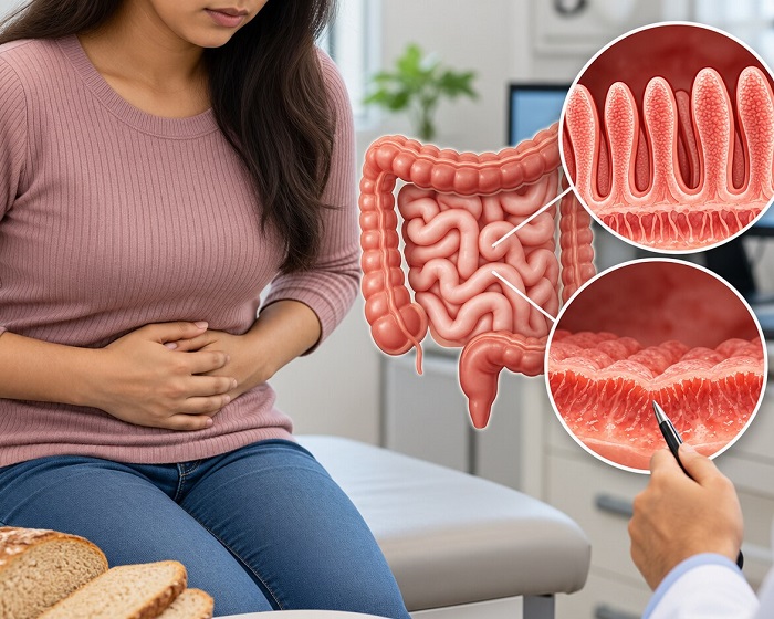
The interior of a healthy small intestine is not a smooth tube. It is lined with thousands of microscopic, finger-like projections called villi, which are further carpeted by even smaller hair-like extensions known as microvilli. Together, they form what gastroenterologists refer to as the brush border.
This anatomical design serves an essential biological purpose: it dramatically expands the absorptive surface area of the intestinal tract, transforming a twenty-foot muscular canal into an expansive landscape roughly the size of a tennis court. Every millisecond of digestion relies on this vast surface area to interface with food, extract nutrients through specialized enterocyte cells, and transport them directly into the bloodstream and lymphatic vessels.
When someone with celiac disease ingests gluten, the immune system misinterprets partially digested gluten fragments (specifically peptides like gliadin) as dangerous foreign invaders. White blood cells infiltrate the intestinal mucosa and release pro-inflammatory cytokines, which inadvertently attack healthy tissue.
Over time, this continuous inflammatory state results in villous atrophy—the flattening, erosion, and eventual blunting of the villi. As these vital structures wear down, the interior of the intestine becomes smooth and scarred. The massive surface area needed for nutrient transfer collapses to a fraction of its normal capacity, crippling the digestive tract’s ability to absorb food regardless of how nutrient-rich an individual’s diet might be.
The Cascading Crisis of Malabsorption
The most immediate and pervasive consequence of villous blunting is generalized malabsorption. Nutrients pass through the digestive tract largely unprocessed and unabsorbed, exiting the body without ever reaching the cells and organs that depend on them.
Because different sections of the small intestine specialize in absorbing distinct nutrients, the location and severity of mucosal damage dictate the specific deficiencies a person develops.
Micronutrient Depletion and Clinical Fallout
The upper sections of the small intestine, primarily the duodenum and upper jejunum, bear the initial brunt of gluten exposure. These regions are responsible for absorbing critical minerals like iron, folate, and calcium.
When these areas erode, the body struggles to maintain basic physiological baselines:
-
Iron Deficiency Anemia: Iron absorption drops precipitously, leading to a chronic, treatment-resistant anemia that causes severe fatigue, pale skin, shortness of breath, and brain fog. Many adults discover they have celiac disease only after traditional oral iron supplements repeatedly fail to raise their ferritin levels.
-
Calcium and Vitamin D Deficits: The body cannot absorb dietary calcium without intact intestinal pathways and active vitamin D. When absorption fails, the body draws calcium out of the skeletal system to maintain blood levels, accelerating bone mineral density loss and predisposing individuals to early-onset osteopenia and severe osteoporosis.
-
Folate and Vitamin B12 Depletion: Damage extending further into the jejunum and ileum compromises the assimilation of B-complex vitamins, which can result in macrocytic anemia, elevated homocysteine levels, peripheral neuropathy, and mood disturbances.
Disrupted Fat Absorption and Secondary Intolerances
The damage does not stop at micronutrients. The breakdown and uptake of dietary fats are severely compromised when villous architecture is lost. Unabsorbed fatty acids travel into the large intestine, leading to steatorrhea—pale, foul-smelling, floating stools that signal severe malabsorption and rapid, involuntary weight loss.
Because fat-soluble vitamins (vitamins A, D, E, and K) require efficient lipid absorption to enter circulation, their levels drop alongside dietary fats. This leads to impaired night vision, poor wound healing, cellular oxidative damage, and compromised blood clotting mechanisms.
Furthermore, enterocytes located at the very tips of healthy villi produce lactase, the digestive enzyme responsible for breaking down the milk sugar lactose. When villous blunting shears off these cell tips, the body completely loses its ability to produce lactase. This induces a secondary lactose intolerance, causing severe gas, cramps, and diarrhea whenever dairy is consumed, even though the underlying problem is gluten-mediated epithelial damage rather than a congenital dairy allergy.
Beyond the Gut: Systemic Repercussions of Barrier Breakdown
The intestinal lining serves a dual purpose: it acts as a selective sponge for nutrients while functioning as a tight physical barrier against toxins, undigested macromolecules, and systemic pathogens.
In active celiac disease, the chronic inflammation damages the tight junctions that seal intestinal cells together, significantly increasing intestinal permeability. When this barrier weakens, immunogenic molecules and environmental antigens leak across the epithelial layer and encounter the systemic bloodstream.
This systemic spillover triggers widespread autoimmune and inflammatory reactions throughout the body:
-
Cutaneous Symptoms: Many patients develop dermatitis herpetiformis, an excruciatingly itchy, blistering skin condition that appears symmetrically on elbows, knees, buttocks, and scalp when circulating antibodies deposit in the dermal tissue.
-
Neurological Impairments: Gluten sensitivity in celiac patients can affect the central nervous system, manifesting as gluten ataxia (loss of motor coordination), chronic migraines, and widespread peripheral neuropathies characterized by tingling or numbness in the hands and feet.
-
Joint and Muscular Inflammation: The circulation of immune complexes frequently produces chronic joint pain and persistent muscular stiffness, often misattributed to fibromyalgia or general arthritis.
The Mucosal Rebuilding Process
The treatment for celiac disease requires an absolute, lifelong commitment to a strict gluten-free diet. There are currently no pharmaceutical remedies that allow someone with celiac disease to consume gluten safely. Even trace contamination—such as crumbs from a shared toaster or shared cooking utensils—is sufficient to reignite the inflammatory cascade and perpetuate mucosal damage.
Eliminating gluten completely halts the autoimmune attack. Without the offending protein triggering immune cells, the mucosal lining begins a methodical process of cellular regeneration. Intestinal stem cells located in the crypts between villi generate fresh, healthy enterocytes that migrate upward, slowly rebuilding the villi and restoring the brush border’s expansive surface area.
Recovery is a gradual process that demands patience and deliberate medical management:
-
Clinical symptoms like diarrhea, acute abdominal pain, and extreme bloating often begin to improve within days or weeks of removing gluten.
-
Complete histological recovery—the full microscopic regrowth and normalization of intestinal villi—typically takes anywhere from several months to a couple of years, particularly in adults who lived with undiagnosed disease for a long time.
-
During the early phases of healing, patients often require targeted nutritional supplementation, occasionally via intramuscular injections or intravenous infusions, to bypass the damaged gut and restore depleted iron, vitamin D, and B12 stores while mucosal architecture recovers.
Celiac disease is ultimately a disease of systemic starvation driven by localized autoimmune destruction. By damaging the delicate landscape of the small intestine, it starves the body of the basic biological building blocks required for cellular health, energy production, and skeletal strength. Understanding the physiological reality of villous atrophy emphasizes why a gluten-free diet is far more than a passing wellness trend for individuals with celiac disease; it is the medical therapy that allows the digestive system to rebuild, absorb nutrients, and restore total-body health.